Menu

Wananga landing Wananga landing
Topic

Equipment

11 December 2023

Outstanding facilities and equipment are critical to the Biomolecular Interaction Centre (BIC)  mission of delivering world-class research in biomolecular interactions at the interface of engineering and science. BIC continues to invest strategically in capital equipment and in our ongoing maintenance. Find out about our equipment.

APPLY NOW

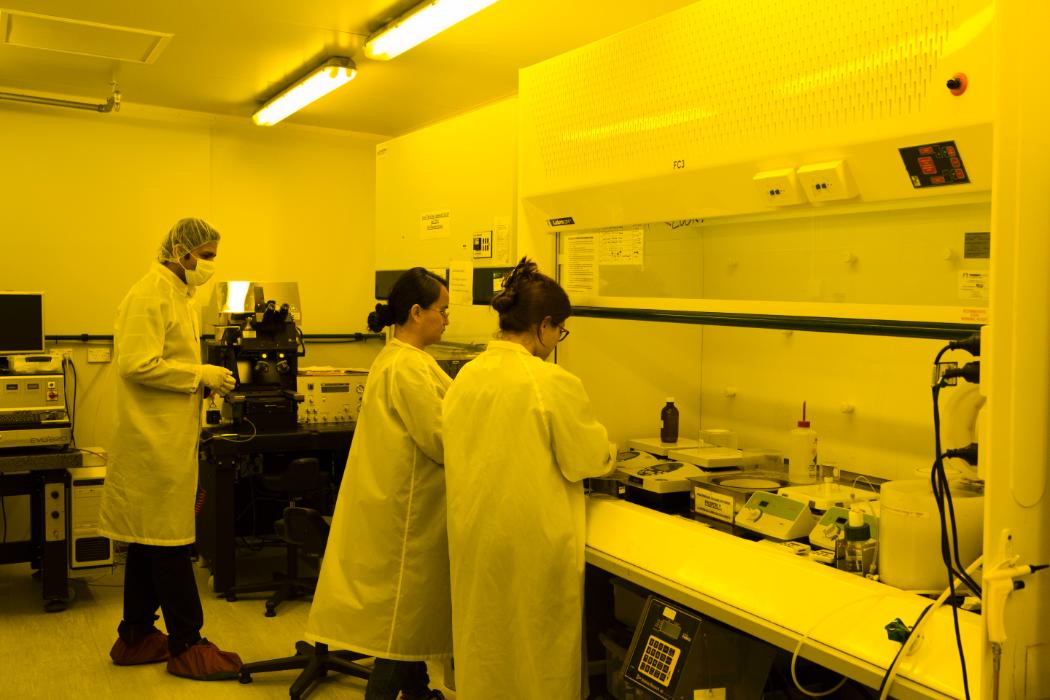
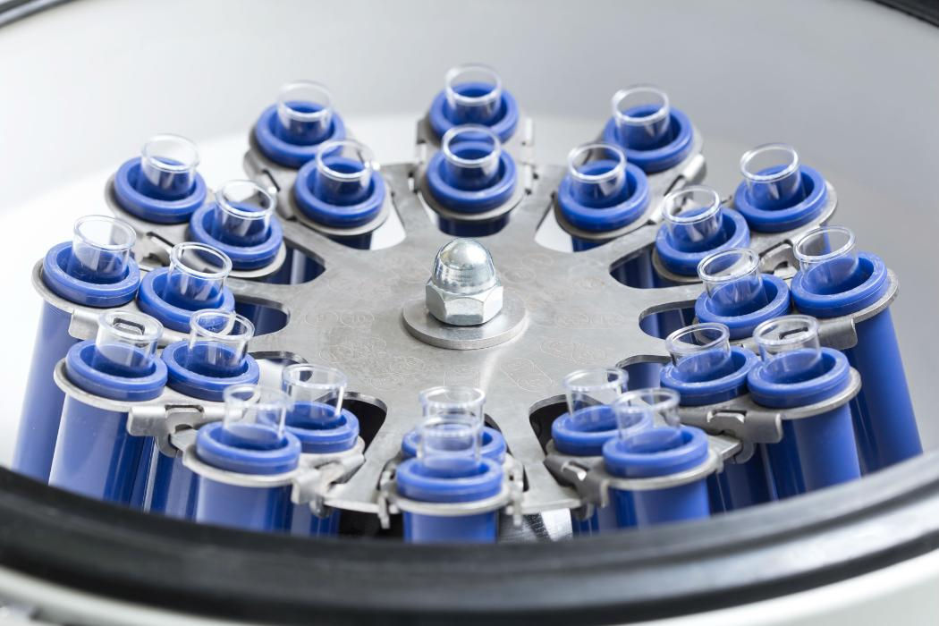
Outstanding facilities and equipment are critical to BIC’s mission of delivering world-class research in biomolecular interactions at the interface of engineering and science. As such, BIC continues to invest strategically in capital equipment and in its ongoing maintenance.

We have a range of advanced technologies to enhance our ability to study biomolecular interactions. If you are interested in learning about or using any of the facilities below, please email biomolecular@canterbury.ac.nz.

The New Zealand Analytical Ultracentrifugation Centre (NZAUC) is the only centre of its kind in New Zealand. The analytical ultracentrifuge allows researchers to probe the molecular mass of biomolecules in solution and to characterise interactions between different biomolecules (e.g. protein-DNA interactions).

Developments in instrumentation and data analysis have greatly increased the potential of the facility to deal with complicated systems, including inorganic crystalline nanoparticles and interactions within cell lysates and food, and to provide information complementary to other techniques, such as surface plasmon resonance (SPR).

The AUC is accessible to outside organisations on a contract or collaborative basis. For more information contact biomolecular@canterbury.ac.nz.

An automated platform used for size and quantitation analysis of proteins, DNA and RNA. It works through microfluidic separation that produces size and electropherogram results for quantitative analysis from pure samples to cell lysates.

Within BIC we have a range of biomolecule purification equipment, including various AKTA systems (Pure, Basic and Express systems) and associated columns and media for any type of purification.  We also have detection systems for absorbance and fluorescence.

Circular Dichroism spectroscopy allows the determination of protein secondary and tertiary structure.  It can also be used to evaluate DNA quadruplex structure.  It is often used to determine the chemical or temperature stability of biomolecules.

A confocal microscope detects the fluorescence emitted when the specimen is scanned by a laser, resulting in high-resolution images and the ability to directly observe dynamic processes in live cells. Cell components can be precisely localised, and accurate three-dimensional information is easily obtained. The behaviour and interactions of molecules can be studied.

Our inverted Leica TCS SP5 confocal microscope is equipped with a full range of lasers from 405 nm to 633 nm, as well as incubation and heating/cooling chambers.

The confocal microscope is accessible to outside organisations on a contract or collaborative basis.

Determine protein folding, domain structure and the effect of solution conditions, mutations, ligand binding and chemical labelling on protein conformation.

A dynamic light scattering system for particle and molecular weight measurements; analysing protein charge and iso-electric point through pH titration; assessing protein aggregation, oligomerisation state and protein-protein binding in a range of solvents; measurements of colloid and emulsion solutions.

This system is able to detect the presence of tagged protein or analyse binding kinetics. Binding events can be viewed in real time, and  kinetic rates measured for association, dissociation and affinity. Assays require only 4 ul of sample, and can be performed in complex mixtures, or even crude solutions.

This analytical facility allows the high-resolution analysis and separation of anions. The detector system used is electrochemical (pulsed amperometric detection, PAD, sometimes also called pulsed electrochemical detection, PED), and is extremely sensitive; significantly more so than typical HPLC detectors.

A wide variety of different columns are available for use with the system, which allow a very broad range of analytical applications.

In addition to anions, as the eluent used is sodium hydroxide, weakly acidic organic compounds, such as reducing sugars are also very amenable to analysis. The ICS system is capable of mono-, oligo- and polysaccharide analysis and profiling, and finds applications in disciplines as disparate as Glycobiology and Food Science.

This instrument is accessible to outside organisations. It requires the purchase of specific columns and eluent. 

The interaction between molecules is often accompanied by a change in thermal energy.  ITC quantifies this change allowing the precise determination of the thermodynamics of a biomolecular interaction. It is useful for protein-protein, protein-peptide, protein-ligand and protein-small molecule interactions.

MicroScale Thermophoresis (MST) is an immobilization-free technology for measuring biomolecular interactions. The MST instrument detects the motion of fluorescent molecules along a microscopic temperature gradient, which reflects changes in the molecular hydration shell, charge, or size. Since one or all of these parameters will change with virtually every binding event, a wide range of biomolecules can be measured, from ions and small molecule fragments to large macromolecular complexes, in small volumes (~20 μl), in a wide range of standard buffers and complex mixtures such as liposomes, detergent, serum, and cell lysates.

The NanoLab contains facilities for semiconductor material processing, nanofabrication, and sensor and microfluidic device development.

The equipment covers most aspects of semiconductor device fabrication, from materials growth and characterisation to device packaging. The principal tools are:

  • Atomic Force Microscope (Digital Instruments Dimension 3100)
  • Deep Reactive Ion Etching (Oxford PlasmaPro 100 Cobra)
  • Electron Beam Lithography (Raith 150)
  • Interference Lithography
  • Mask Writing (Heidelberg uPG101)
  • Micromilling (CNC Mini- Mill/GX)
  • Nanoimprint Lithography (EVG)
  • Optical Lithography (Suss MA-6)
  • Optical Microscopy (Olympus BX30 with digital image capture)
  • Photonic Professional GT2 two-photon polymerisation 3D printer
  • Plasma Ashing (Tergeo plasma cleaner)
  • Semiconductor Device Characterisation (HP 4155A Parameter Analyser)
  • Surface Profilometer (Profilm3D)
  • Thin Film Deposition (Edwards AUTO 500, Temescal FC-1800 and Mist CVD)
  • Wire Bonding (Kulicke & Soffa 4500)

The facility is accessible to external organisations on a contract or collaborative basis.

Equipped with a Fluorescence and Florescence anisotropy detector and heating to 60 degrees.

Set up protein crystallisation trials easily and effectively.  The drop sizes are usually 200—400 nL and the usual setup is a 96 well plate.

Combines a thermal cycler and optical reaction module for singleplex and multiplex detection of fluorophores. The system features thermal gradient functionality and automation capabilities. In addition to real-time polymerase chain reactions (PCR), the instrument also offers rapid and simple protein stability screens by thermal melts.

Scattering of visible light can be used to determine the size and overall dimensions of macromolecules in solution. Absolute molecular weight can then be calculated directly from the light scatter, but this requires accurate measurement of protein concentration. To achieve this the Viscotek has detectors for refractive index and UV. Additionally, the Viscotek has a viscometer, which allows determination of molecular size, conformation, and structure.

A protein interaction array system that enables label-free quantitative analysis of biomolecular interactions in real time using surface plasmon resonance (SPR) technology. The interactions are monitored over time by flowing an analyte in a microfluidic channel over a ligand immobilized on a sensor chip and detecting the binding of the analyte to the ligand by measuring changes in the propagation of electromagnetic waves at the sensor surface.

Can analyse up to 36 different protein, peptide, DNA and small molecule interactions in a single run.

Utilised to monitor the speed of biomolecular reactions. Equipped with multicell block and temperature control.

The Bruker 600 MHz with a cooled sampler allows for automated measurement of a large number of biological samples. This is an optimal set up for the measurement of clinical samples and is currently the only one of its kind in Aotearoa New Zealand. Our instrument also allows for the analysis of large biological molecules sich as proteins, nucleic acids and carbohydrates.

Privacy Preferences

By clicking "Accept All Cookies", you agree to the storing of cookies on your device to enhance site navigation, analyse site usage, and assist in our marketing efforts.