

In the western world stroke is the third largest cause of death, following heart disease and all forms of cancer, and the largest cause of serious long-term disability. The monetary costs associated with stroke care are considerable and it is projected that if the incidence of stroke continues to increase at the same rate it will more than triple in the next 30 years. There is therefore a great importance on the understanding of factors that increase the risk of stroke and hence how blood is distributed throughout the brain.
The current research is focused on a ring-like arterial structure located in the base of the brain, known as the Circle of Willis.
The function of the Circle of Willis is to distribute oxygenated blood entering through the afferent basilar and internal carotid arteries, to the cerebral territories through three major pairs of efferent arteries known as the anterior, middle and posterior cerebral arteries. The circle is of particular importance because it allows for blood to be re-routed through the anterior and posterior communicating arteries in order to maintain oxygen supply to all of the cerebral territories, should the blood supply through any of the afferent arteries be reduced. Maintaining blood supply is important since even though the brain only comprises approximately 2% of the body’s total mass, it requires approximately 20% of the blood output from the heart, and if the brain is starved of this supply for more than a few minutes brain cells become permanently damaged.
Among the general population, approximately 50% have a complete circle of Willis, where among a multitude of possible anatomical variations, vessels which are greatly reduced in diameter or even completely absent are common, either of which reduces the degree to which blood may be re-routed through the circle. While an individual with one of these variations may, under normal circumstances, suffer no ill effects, there are certain pathological conditions, a build-up of atherosclerotic plaque in the afferent arteries, which can present a risk to the person’s health and the possibility of suffering an ischaemic stroke, when compounded with the effects of an absent vessel.
An advanced 3D computer model has been developed from Magnetic Resonance Imaging (MRI) data and uses a technique known as Computational Fluid Dynamics (CFD) to model the blood flow throughout the circle of Willis.pathlines The simulation results give the response of the circle to different pathological conditions, including the combined effects variations in cerebral-vasculature with atherosclerotic plaque build-up. The long-term goal of the project involves the development of a clinical diagnostic tool for automatically recreating a 3D model of an individual’s cerebral vasculature, from an MRI scan and using CFD to predict the likelihood of stroke in the short term, or the risk associated with various surgical procedures such as carotid endarterectomy.
This project is under the supervision of Professor Tim David. Postgraduate students working on the project include Steve Moore, Samara Alzaidi, Kate Moorhead, and Jade Arnold. This work is being conducted in cooperation with researchers from NASA Ames Research Center.
Chronic kidney disease includes conditions that damage the kidneys and decrease their ability to maintain homeostasis in the body. As kidney disease progresses, waste products can rise to high levels in the blood and cause illness and complications like high blood pressure, anaemia, weak bones, poor nutritional health, and nerve damage. Also, kidney disease increases the risk of developing heart and blood vessel disease, deterioration which can occur over a long period of time. Chronic kidney disease may be caused by diabetes, high blood pressure and other disorders. When kidney disease progresses, it may eventually lead to kidney failure, which requires dialysis or a kidney transplant to survive. There is therefore great emphasis placed on the understanding of factors that increase the risk of kidney disease and how blood is distributed and autoregulated throughout the kidney, enabling proper filtration.
The current research is focused on modelling the kidney on multiple levels, from the functionality of the whole organ down to autoregulation in the nephron, the filtration unit of the kidney. There are approximately one million nephrons in each human kidney, each being an independent entity capable of producing urine via plasma filtration.
The filtration and excretion of metabolic waste is a major function of the kidney that is closely related to its' other important task, maintaining desired concentrations of key constituents in bodily fluids, namely sodium. The filtration, reabsorption, and excretion process of the nephron is elaborate, and there is a large amount of evidence that suggests chaotic interactions between neighbouring nephrons, which adds another degree of complexity to the model. In gaining a greater understanding of the mechanisms governing this balance, we hope to develop novel diagnostic and treatment approaches that are more effective than current medical practices.
This project is under the supervision of Professor Tim David. This work is being conducted in cooperation with Dr Mike Plank and Dr Alex James of the Mathematics department and Professor Zoltan Endre, a nephrologist from the Christchurch School of Medicine and Health Sciences.
Spinal Cord Injury (SCI) is one of the most complicated injuries the human body can sustain. Simply surviving a spinal cord accident is a miracle in itself. However unlike most injuries, damage to the spine is irreparable and the person with the injury must prepare themselves for a dramatic change in lifestyle.
Tetraplegia is a type SCI located in the neck region of the spine and results in paralysis of the upper and lower body. Due to the decrease in functionality of the upper extremities, such as the arms and hands, it becomes difficult to perform simple tasks such as writing with a pen or opening a door. These everyday activities which are necessary to function on a day-to-day basis are commonly referred to as Activities of Daily Living (ADL).
C5 tetraplegia is the most common neurological level of legion (14.8%), affecting generally young males. The greatest disadvantage for people with C5 tetraplegia is the loss of hand function and according to many researchers 75% of persons with tetraplegia would rather regain hand function than any other function that they have lost, including bowel function, bladder function, sexual function, and even the ability to walk.
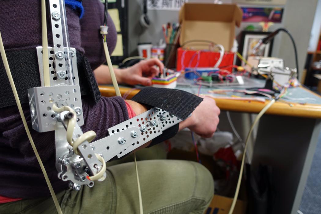
Professor Alastair G Rothwell, Head of Department for Christchurch school of medicine and health sciences proposed a solution to provide a person with C5 tetraplegia the means to grasp and manipulate objects. His proposal was that if a mechanical device could provide wrist control, then combined with his surgery of the thumb, he could provide a person with C5 tetraplegia the ability to perform a key pinch grip. This grip would enable a person with C5 tetraplegia to perform activities of daily living and increase their independence and quality of life.
The research focused on the concept development phase of the design process. The aim was to create a solution which was not only functional, but also acceptable to the user. The research involved generating and selecting the design requirements specifications and developing various concept solutions. This information was then used to develop the optimal concept design.
The final concept holds the wrist in extension by a spring which connects between the hand and wrist brace. When the user wishes to release the object, they increase the force in the cable running underneath the arm. The increase in force flexes the wrist downwards and the object drops out of the hand. The cable running underneath the arm is controlled by a combination of shoulder abduction and flexion as well as protraction and retraction of the auxiliary shoulder.
The final concept can be hidden under loose clothing making the user appear ‘normal' to avoid unnecessary attention.
The project is under the supervision of Dr Shayne Gooch, and co-supervised with Professor Harry McCallion and Professor Timothy David. Other researchers who have been involved with this project include Lan LeNgoc, Marcus King, Julian Verkaaik, and Jennifer Dunn. The project was funded with a scholarship from Industrial Research Limited (IRL) and also generously supported by the Artificial Limb Centre based at Burwood Hospital.
Cardiovascular Systems Modelling and Model-Based Diagnosis
Professor Chase is the leader of this research area if you wish to receive more information
This project uses models of the cardiovascular system (CVS). We are modelling and system identifying a complex heart and circulation model for use in critical care for advanced diagnosis and therapy decision support using measurements from catheters and sensors already commonly used in critically ill patients - i.e. no added cost or technology required.
These models are applied to sort a wealth of clinical data and develop patient-specific diagnoses from otherwise limited and confusing data, which entails using the stream of numbers that are output to identify model parameters. Identified model parameters thus take this data and create a picture of the physiological system status for the patient. The outcome is thus a patient-specific model on which different treatments can be applied/tested to optimise therapy.
We are just entering a more clinically oriented phase that will see a mix of modelling and clinical development. Prior to this we have done extensive animal trials to validate the system with partners at the University of Liege in Belgium.
This area also has some need for mechatronics oriented research work developing better data gathering systems and methods, as well as real-time data analysis if that is an area of interest.
ARDS Lung Mechanics for Optimizing Ventilator Treatment
Professor Chase is the leader of this research area if you wish to receive more information
This project develops a model of mechanics of passively breathing lung, as in case of an Acute Respiratory Distress Syndrome (ARDS) patient under mechanical ventilation. The model is based on the simplified physiology of actual lungs and it uses relatively new concept, where the majority of volume change occurs by recruitment and collapse of alveoli units. The model captures the dynamic characteristics of lungs through Unit Compliance Curve and Threshold Opening and Closing Pressure of lung units. The ultimate goal of this project is to develop a model based decision-support tool for clinical situation, where the model simulates the patient and condition specific lung from a few clinical data and determine the updated optimal setting for the ventilator.
Modelling and Control of the Insulin-Glucose Metabolic System to control Hyperglycemia in Intensive Care and Diabetes Mellitus patients
Professor Chase is the leader of this research area if you wish to receive more information
Diabetes is a widespread problem reaching epidemic proportions around the world. The high blood glucose concentrations, or hyperglycaemia, resulting from Diabetes is a cause for further complications and a higher mortality risk for the affected patients. Intensive care patients are often hyperglycaemic due to the stress of their condition. A tight control of blood glucose in this population has shown to drastically reduce their mortality rate.
The body utilizes insulin to remove glucose from the blood plasma. However, patients with hyperglycaemia have an impaired endogenous insulin metabolism, which can not cope with the task of supplying the right amount of insulin to maintain a normal range of blood glucose concentration. Inter- and intra-individual variability in insulin sensitivity, as well as variability in patient condition, makes it very difficult to find a general, one-size-fits-most solution to the highly nonlinear problem of optimising insulin dosing and glycaemic control.
This project deals with:
- Automating the monitoring and dosing of insulin for intensive care and diabetes patients, and applying advanced modelling and adaptive control design for the automation of insulin infusion. This area includes theoretical work in simulating and optimizing the trial protocols as well as practical implementation of these in proof of concept clinical trials.
- Improving current models to improve the predictive ability of the system and account for greater physiological variations and effects.
- Implementation of emerging continuous glucose sensors into closed loop systems to reduce human interaction. Problems arising due to sensor lag and errors have to be dealt with by applying appropriate filtering and control techniques.
- Development of a low-intensity model-based test to quantify of insulin resistance for the broad population, focusing on clinical practicability and higher accuracy than current methods.
This multi-disciplinary project is carried out in cooperation with the following institutions:
- The Christchurch Hospital ICU
- The Department of Mathematics and Statistics
- The Edgar National Centre for Diabetes Research, Dunedin, NZ
- The Lipids and Diabetes Research Group at Christchurch Hospital
Initial proof-of-concept clinical trials are carried out in the Christchurch Hospital ICU. The current control protocol involves constant monitoring of patient blood glucose levels and reviewing of patient-specific parameters that govern the blood glucose variability. Through theses step thus the insulin requirement can be determined and administered.
Sedation-Agitation Sensor for Intubated Patients in the ICU
Professor Chase is the leader of this research area if you wish to receive more information.
Intensive care unit (ICU) patients are often intubated to help them breathe, and sedated to minimise pain and agitation from the intubation as well as other injuries. Patients that are not sedated enough often become agitated and try to remove the breathing tube causing distress and anxiety that are difficult to control without unnecessary extra sedation.
The goal of this project is to:
- Create a sensor array to measure patient motion with existing sensor technology
- Correlate and quantify patient motion to existing qualitative agitation scales
The basic premise is that patient motion, and other metrics, are directly correlated to patient agitation. Current measures of patient agitation are qualitative relying on medical staff to make periodic, subjective judgments.The application of modern sensor and signal processing technology presents the opportunity to gather more data and apply it to create a qualitative, far more precise, determination of patient agitation. Success would enable better sedation-agitation modelling as well as a more quantified approach to controlling sedation processes. This project is run in conjunction with Dr Geoff Shaw, M.D. a research anesthesiologist with the Christchurch Hospital and the Otago School of Medicine.
Modelling and Control of the Sedation-Agitation Curve in ICU Patients
Professor Chase is the leader of this research area if you wish to receive more information.
Intensive care unit (ICU) patients that are not sedated enough often become agitated and try to remove the breathing tube causing distress and anxiety that are difficult to control without extra sedation. Conversely, over, or heavily, sedated patients take significantly longer returning to a conscious state, adding significant cost and time to their hospital stay as well as additional risk due to over-sedation.
The primary problem is twofold:
- Lack of an adequate model relating agitation and sedation
- Inability of shrinking nursing staffs to consistently understand, dose and treat sedated patients with the minimum necessary sedation, i.e. lack of automatic control
This project looks at addressing these two problems. The first part creates a quantifiable sedation-agitation model suitable to covering the majority of patient behaviours in terms of relating sedative concentration to qualitative level of sedation and a quantified level of measured agitation. The second part examines applying control systems technology to this system to obtain more robust and consistent results, and to achieve more minimal levels of sedation to minimize ICU stays and healthcare cost. This project is also run in conjunction with Dr Geoff Shaw, a research anesthesiologist with the Christchurch Hospital and the Otago School of Medicine.
An interdisciplinary team is investigating a novel breast cancer screening method aimed at imaging elastic properties within soft tissue. This work involves full-time faculty, staff, post-graduate and undergraduate researchers as well as research scientists from industry.
Digital Image Based Elastic Property Assessment Elastic property imaging is undergoing rapid development across the globe as its potential for effective cancer screening and diagnosis is better understood. Most methods being currently developed are based on Magnetic Resonance or Ultrasound modalities, making them either too costly or too limited in their field of view to be practical screening mechanisms. This project aims to develop an elastic property imaging technique targeted as an effective screening method, meaning it must be relatively low cost and yet provide a complete field of view for analysis of the full breast.
The fundamental concept behind this technique is the reconstruction of an elastic property description of the breast volume based on motion information obtained at the breast surface by 3D digital imaging techniques. This type of imaging is referred to as a tomographic technique and is similar in concept to an X-ray CT scan except for differences in the physics of the wave propagation that the different systems must account for. X-ray CT scanning involves the measurement of x-ray particle wave patterns at the tissue surface while the proposed elasto-tomographic system would rely on the measurement of mechanical wave patterns at the surface.
The specific project involves three main components:
- The development of the 3D digital imaging system for capturing the surface motion of the breast.
- The development of the computational algorithms to convert these motions into descriptions of the internal elastic properties of the breast.
- The development of the mechatronic hardware required to generate the surface motions on the breast and mobilize the digital imaging system.
From Mechanical Engineering Professor Geoff Chase has a broad range of expertise in digital control and actuation. This work is being conducted in cooperation with researchers from the Eastman Kodak Company in Rochester New York, a world leader in the medical imaging industry.
The focus of this research is to understand and model the effects of arterial geometry on nucleotide and endothelium cellular processes. The project brings together a group of researchers, from a diverse range of backgrounds, in the Departments of Engineering, Mathematics and Biological Science.
Nucleotides and the Endothelium
Adenine nucleotides such as adenosine triphosphate (ATP) and diphosphate (ADP) have been shown to be important modulators of vascular tone (the muscle tension in the arterial wall). These nucleotides react with receptors on the endothelial cells, and control arterial geometry (vasodilation) via the production of Endothelium-derived relaxing factor (EDRF). Nucleotides can be present in flowing blood as a result of the activation of platelets and from vessel wall damage. At the endothelial surface the agonist ATP undergoes degradation via the following reaction mechanism: ATP → ADP → AMP → adenosine, where the latter two have been observed to be in low concentration due to slow reaction rate. The concentration profile of nucleotides at the surface of the endothelial cells is dependent on the velocity profile of the blood which brings ATP from upstream. ATP needs to be continuously transported to the surface of the endothelium in order to regulate responses; this transport to the surface is brought about by convective and diffusive processes. Haemodynamic shear stress, the tangential force exerted on the endothelial cells by the flowing blood, evokes a number of responses by the endothelial cells; in particular it has been demonstrated that endothelial cells respond to fluid shear stress by increasing cytosolic free Ca2+ concentration.
Models in Production
Previously, mathematical models have been developed to evaluate the concentration of nucleotides at the endothelium. Specifically, the model used in this research can take any “well behaved” wall shear stress function, which is related to vessel geometry, and evaluate the nucleotide concentration at the endothelium.
The main focus of this research, in terms of nucleotide concentration, is looking at the concentration variation in an arterial bifurcation; this geometry is important as it is known to have numerous effects on the health of the cardiovascular system ie. atheroma occurs preferentially where the wall shear stress is low or spatially vary, which is associated with this geometry.
Numerical models, for similar flow scenarios as the analytical, are also important; these models provide useful comparisons and validation of the analytical model results. Additionally, certain flow scenarios, such as a 2D arterial bifurcation, have wall shear stress functions that cannot to be obtained analytically, so the wall shear stress must be taken from numerical simulations; this can then be used to evaluate nucleotide concentration at the endothelium.
Numerical methods are foundational to most complex engineering applications. However, numerical methods are typically poorly applied in biomedical applications. This research stream aims to improve current numerical and analytical methods in bioengineering to make tangible improvements in diagnosis, prediction and treatment.
Fields of research within numerical methods for bioengineering:
- Implementation and improvement of parameter identification methods in pulmonary mechanics
- Development and application of novel methods of forward simulation for the acceleration of simulation and improved robustness of the inverse problem
- Alternative methods of the inverse problem when dealing with large data sets of dynamic data
- Structural and practical model identifiability analyses for improved protocol design and information yield
- Model observability in the presence of ‘grey noise’ and poorly sampled data
- Dynamic signal analysis using Bayesian approaches.
- Novel approaches in fluid dynamics and thermodynamics.
Please contact Dr Paul Docherty for further information.
In the last two decades, with the combined advancement of biological sciences and materials engineering, the new field of tissue engineering has evolved. With the recent rapid developments in these converging fields, the potential market worldwide for tissue-engineered products is estimated at nearly €100 billion per year. Tissue engineering (TE) strategies which aim to combine patient’s own cells, biodegradable scaffolds and growth factors may offer considerable advantages over current surgical interventions used to repair or regenerate damaged tissues following trauma or disease.
Tissues have complex three-dimensional (3D) geometries and highly organised internal architectures which cannot be simply emulated by cells maintained in two-dimensions. Porous scaffolds are central to tissue engineering strategies because they provide a 3D framework for delivering reparative cells or regenerative factors in an organised manner to repair or regenerate damaged tissues. While these tissue engineering strategies have shown promising results, a number of challenges need to be overcome before these strategies are widely accepted clinically.
Our BioMATE group has research focused in the following areas:
- Biomaterials Engineering
- Design and processing of polymeric, ceramic, metal alloys materials
- Tissue Engineering and Regenerative Medicine
- Cartilage
- Bone
- Neural/spinal cord
- Advanced scaffold design and bio-manufacturing
- Solid free-form fabrication of porous scaffolds
- Mechanical analysis
- Pore architecture optimisation and modelling
- Novel metal alloys for bone interfacing implants
- Electrospinning of biopolymer nanofibres
- Orthopaedic biomechanics
- Total joint replacement
- Medical device design
UC collaborations
- Atherosclerosis (free radicals and anti-oxidants) applied to vascular surgery at CHMEDS
- Biotechnology
- Electrical Engineering
- Biomedical Signal Processing applied to EEG and ECG
- Electrobiotechnology applied to the use of electromagnetic signals in biology and medicine, particularly with respect to high voltage cellular effects
- Biomathematics applied to cell calcium dynamics
- Optimisation (of cardiovascular systems)
- Medical and Clinical Statistics and statistical DSP modelling
- Bioinformatics, genomic research, health environmetrics
- Biological Computational Fluid Dynamics applied to cardiovascular blood flow, blood flow in the brain and Cellular motion in the eye
- Biological Control Theory applied to non-invasive imaging technologies for breast cancer and insulin control and monitoring. Research and development of the latest agitation-sensor and automated drug infusion systems for ICU Patients
- Orthopaedic implant design and development
- MR imaging with particular reference to cerebal perfusion (e.g. ASL and BOLD protocols)
External collaborators
- Neurology applied to blood flow in the cerebro-vasculature
- Radiology applied to cerebro-vascular imaging
- Orthopaedic implant design
- Thrombolysis protocols
- Renal autoregulation; renal failure
- Non-invasive breast cancer screening
- Insulin drug infusion
- Visualisation Techniques applied to medical imaging and numerical simulations
- Cerebro-vascular blood flow in micro-gravity
- Numerical models of blood flow and mass transfer and muscle microstructure. (Dr Spencer J Sherwin)
- Visualisation of arterial geometry and grid computing (Professor Ken Brodie)
- Thrombolysis protocols, eye fluid dynamics
- Bioinformatics
